Infectious laryngotracheitis is a viral disease caused by Herpes group of virus infection affecting layer and broiler flocks resulting in dyspnoea and expectoration of bloody mucus in its most severe form, with high morbidity and mortality rates. It is found in all parts of the world but in India, there exists scant reference on its occurrence. The present report depicts its persistence on a broiler farm over several successive cycles, and how a group of veterinary experts was able to identify the cause and bring the disease under control.
The affected broiler farm has a capacity of more than 50,000 birds, and each weekly cycle introduces a further 2,000 day-old broilers, which are marketed at an average of 40 days of age.
According to the farm supervisors, morbidity and mortality in up to 80% of the flock started around one year previously and attempts to control the disease had been unsuccessful. It had been found that the cause of early mortality in the first two weeks of life was due to urolithiasis, characterised by distended ureters containing uroliths and visceral gout. Of the survivors, 90% demonstrated dyspnoea and sound of moist rales later in life, and 50-60% were diagnosed with complicated chronic respiratory disease' by the farm supervisor.
Tracing the urolithiasis
Various possible causes of urolithiasis in the young chicks were proposed. Among these was dehydration in hatchers, which was checked by the hatchery manager but ruled out. The workers in the brooder house paid particular attention to ventilation, over-crowding, brooder temperature and an adequate supply of drinking water with electrolytes. In case of the possibility of a nephrosis-inducing form of infectious bronchitis virus (IBV), a Massachusetts serotype (mass type) vaccine suitable for day-old chicks by intra-ocular drop vaccination was administered to several successive flocks. Lastly, the protein content of the pre-starter feed was reduced to avoid additional pressure on the kidneys.
Unfortunately, none of these measures made a significant impact on the urolithiasis problem.
Investigating the respiratory disease
Considering the possibility of mycoplasmosis, a programme was initiated to control Mycoplasma gallisepticum and any secondary bacterial complications; a suitable control programme was adopted for several successive flocks. Strict water sanitation was strictly adopted. Again, no improvement in the situation was observed.
Identifying the effects of the ILT virus
It is noteworthy that after several months, a new employee carried out a post mortem and noted white mucus-like material in the wind pipe, having a congested appearance' in a telephone conversation to the veterinary experts. This introduced the possibility of the cause being infectious laryngotracheitis (ILT). Photographs were taken at the farm, and materials collected over the following days. For histopathological examination, specimens are submitted to a private medical laboratory.
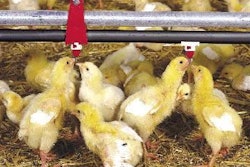
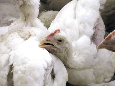
On the farm, an affected 4-week-old broiler was seen with its slightly swollen face and mild cyanosis (Figure 1). There were few cases of severe respiratory distress, gasping with an open beak. The expectoration of mucus and blood-stained exudates, which are classical symptoms of ILT, had been overlooked, confused with signs of coccidiosis.
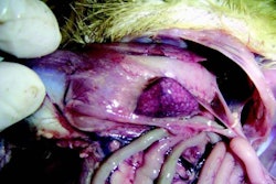
Gross lesions were confined to the upper respiratory tract and most common is haemorrhagic tracheitis (Figure 2). In some cases, caseous, diphtheritic exudates were found in the trachea and as plugs in the laryngeal region.
Figure 4 shows the presence of intra-nuclear inclusions in the epithelial cells that have been sloughed from the inflamed mucus membrane of the trachea. The eosinophilic nature of the inclusions would have been more obvious using other staining methods but these were not offered at the chosen laboratory. However margination of chromatin was clearly visible in the nuclei containing characteristic intra-nuclear inclusions of ILT. Autolytic changes may have taken place in desquamated cells, destroying the cytoplasmic structure. Inclusion bodies were found in only one of the sections.
Figure 5 shows the deposition of uric acid on the internal organs of a 4-week-old broiler (visceral gout), which had hemorrhagic tracheitis.
In his book published in 1969, Text Book on Diseases of Poultry Including Cage Birds and Pigeons, the Australian diagnostician, Dr T.G. Hungerford, states, "When a natural outbreak of laryngotracheitis occurs, e.g. in a broiler plant where uraemia is present, the combined diseases may virtually wipe the unit out. This has occurred on a number of occasions in our experience."
This appears to have happened in the present case, where aggravation of uraemia in an ILT-infected flock was due to disturbed functioning of kidneys relating to the respiratory trouble.
Result of the study has brought into light a fact that apart from existing commonly found diseases in a country, other ailments may be present which on close and open-minded search may be found by diagnosticians.
Control of the disease
The farm management was advised to adopt strict hygiene and biosecurity measures in future, as well as to adopt an all-in, all-out' policy. Vaccination against ILT could not be recommended as no such vaccine is available in India.