Diagnostic test for poult enteritis complex
A new, highly sensitive diagnostic test to detect viruses associated with poult enteritis complex, or PEC, has been developed by Agricultural Research Service scientists in Athens, Georgia, USA. This disease of young turkeys causes diarrhoea, poor weight gain and, in some cases, high mortality. Microbiologists Erica Spackman and Darrell Kapczynski developed the test in a format that allows the detection of several types of viruses at one time.
The test relies on a molecular technique called real-time reverse transcription- polymerase chain reaction (RRT-PCR), which is a highly sensitive and specific method for detecting viral RNA. Current diagnostic methods for PEC-associated viruses have limitations because of poor analytic specificity and sensitivity.
The researchers inoculated turkey poults with each of the PEC-associated viruses and later collected intestinal tissue samples and cloacal swabs from the experimentally infected birds. The RRT-PCR test showed high sensitivity, accurately detecting the target viruses in both the tissues and swabs.
While intestinal samples have previously been essential to making a definitive PEC diagnosis, the researchers found that cloacal samples – which are easier to collect and process – were just as suitable for testing. Using them will not only save time and money, it will also eliminate the need to euthanise birds for sampling.
— ARS news service
Botulism warning
Scottish Agricultural Colleges (SAC) Veterinary Services is urging farmers to be aware of the risks of botulism in cattle that are in contact with broiler litter or animal feed contaminated with wild bird or rodent carcasses. Although rare in Scotland, four cases of botulism in cattle have been investigated by SAC recently. In two of these outbreaks, contact with broiler litter was the suspected source of infection. In the other two cases, it appeared to come from home-mixed feed or silage that had become contaminated with carcass material.
The four cases in Scotland follow reports of an increase, in recent years, in the number of cases of botulism in cattle and sheep in other parts of the UK. In some cases, mortality has been high: 88 animals in a flock of 230 ewes either died or had to be destroyed in one case in England.
— SAC Veterinary Services, UK
Research holds clue to fishmeal’s X-factor
New research in the International Journal of Poultry Science has confirmed the performance-boosting effects of fishmeal in commercial broiler diets, and may go some way to explaining the ‘unidentified growth factor’ associated with this protein source.
Feeding trials conducted at Kurdistan University in Iran showed that daily weight gain and feed intake increased significantly with fishmeal inclusion throughout the 42-day programme.
The beneficial effects of fishmeal on broiler performance became most evident at higher inclusion rates and during the mid-points of the growth period (21-32 days), mainly through stimulation of increased feed intake. At the end of the trials, the average bodyweight of broiler chicks receiving higher levels of fishmeal was more than 9% greater than those fed on a diet without fishmeal.
“This latest research clearly highlights the role of fishmeal in stimulating feed intake. In other words, feed containing fishmeal is more palatable to livestock, and this factor alone results in improved performance and growth,” commented Helge Korsager, chairman of the Fishmeal Information Network.
— Fishmeal Information Network
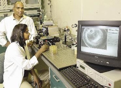
Shedding new light on bacteria detection
Researchers at Purdue University in the USA have developed a low-cost system that analyses scattered laser light to identify bacteria for applications in medicine, food processing and homeland security at one-tenth the cost of conventional technologies.
The technique is called Bacteria Rapid Detection Using Optical Scattering Technology, and it works by shining a laser through a Petri dish containing bacterial colonies growing in a nutrient medium. It does not require biochemical staining, DNA analysis or other types of manipulation.
Particles of light (photons) bounce off the colony, and the pattern of scattered light is projected onto a screen behind the Petri dish. This ‘light-scatter pattern’ is recorded with a digital camera and analysed with sophisticated software to identify the types of bacteria growing in colonies.
The work was initiated by Professors Arun Bhunia and E. Daniel Hirleman. Their paper has been published in the Journal of Biomedical Optics.
“We adapted some ideas from that research to build a scatterometer for food safety, and now we’re using the second generation of that instrument,” Professor Hirleman said. A major motivation for the research is to reduce the time it takes for industry to identify harmful organisms in food processing. Scientists in food-processing plants routinely grow cultures to test for dangerous pathogens.
A mass-produced system based on the new technology would consist of inexpensive, off-the-shelf hardware, such as red lasers and low-resolution digital cameras available at consumer electronics stores, and likely would cost less than US$1000, commented Professor Hirleman.
The researchers have used the new system to classify six species of Listeria, only one of which is a dangerous food-borne pathogen for humans. It was also able to identify accurately other types of bacterial colonies, including Salmonella and E. coli.
“We were able to classify bacterial colonies with greater than a 90% probability of being correct, which is as good as you could do with equipment costing more than $100,000,” said Bartek Rajwa, a staff scientist also working on the project. “And, unlike conventional systems, our method is 100% non-invasive, which means we can carry out the procedure without staining, manipulating or killing the biological samples.
A provisional patent has been filed for the data-processing technique and a full patent application has been filed on the underlying light-scattering technology.
— Purdue University News